 Oculista Bolzano
Why checks matter when vision seems normal
Glaucoma is a group of conditions that damage the optic nerve. Common chronic forms can progress without pain or an obvious change in central vision. Reading the chart well does not exclude early damage.
Eye pressure matters, but cannot diagnose or rule out glaucoma by itself. Pressure may be high without documented damage, while glaucoma can occur within the usual pressure range. Assessment brings together the optic nerve, corneal thickness, drainage angle, visual field and risk factors.
Family history, age and previous findings affect follow-up. Bring earlier results and mention steroid medicines, including eye drops. A suddenly painful red eye with blurred vision, halos, nausea or vomiting may indicate acute angle closure and needs urgent care.
What OCT RNFL measures
OCT measures structures including the retinal nerve fibre layer, or RNFL, and ganglion-cell layers. It helps identify changes and monitor them over time. This quick, non-contact examination still needs an adequate-quality scan.
A red area on the printout is not automatically glaucoma. High myopia, anatomical variation, segmentation errors and poor images can affect the result. A normal-looking report also needs interpretation alongside other findings.
Comparison with earlier scans, ideally using comparable equipment and protocols, is often more useful than one image. The clinician checks technical quality before attributing a change to disease.
Visual fields: preparing for the test
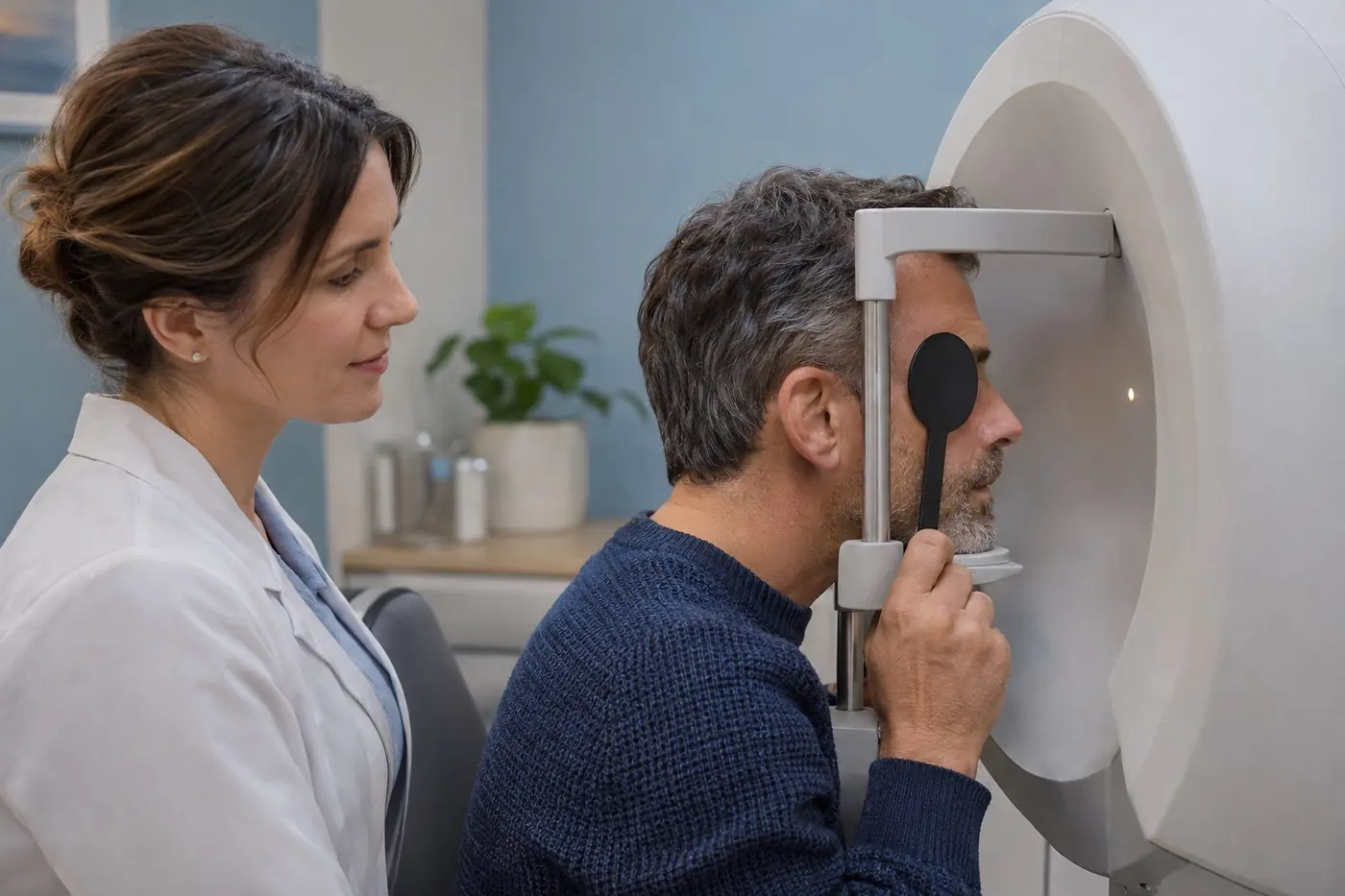
Visual-field testing measures function: you respond to small lights while looking at a fixed target. It complements OCT, which mainly describes structure. The tests answer different questions and are not interchangeable.
It is normal not to see every light. Do not follow lights with your eyes or try to guess. Mention an uncomfortable position, a fogged test lens or the need for a break.
Fatigue, learning and fixation problems can influence the result. An uncertain first test may need repeating; that alone does not establish worsening glaucoma. Progression is assessed through consistent findings and their evolution.
Treatment and follow-up intervals
Treatment lowers eye pressure to reduce the risk of further damage. It does not restore lost nerve tissue. Options include drops, selective laser trabeculoplasty for suitable forms and surgery. The choice depends on glaucoma type, severity and response.
Target pressure and appointment intervals are individual. New diagnoses, progression or treatment changes may require closer monitoring than a stable situation. A six-month interval is not a universal rule.
Do not stop drops because you can see well. Persistent burning, redness, breathing difficulties or other adverse effects need medical advice. Bring a medication list and ask for your drop technique to be checked if necessary.
Sources · European Glaucoma Society: OCT imaging at 6-month intervals · European Glaucoma Society Guidelines, 6th Edition
All sources and further reading (2)
Illustrative images from the website’s image library.
- AI-generated illustrative image
- AI-generated illustrative image