 Oculista Bolzano

Recognising a new difference
A red package may look vivid with one eye but washed out or dimmer with the other. Other colours may change too, or the main impression may be that one eye sees the scene with its brightness turned down. These are useful descriptions even without a medical name.
Contact an ophthalmologist today if the change developed over recent days or is worsening. Mention pain when looking sideways, a central patch or reduced clarity. Abrupt deterioration or blackout requires immediate assessment even when brief. Call 112 for major loss or associated speech difficulty, facial drooping or one-sided weakness.
A mild, longstanding stable difference can be discussed at a planned appointment, or assessed routinely if never examined. You may only have noticed it because you happened to cover the better eye. If onset is uncertain, give the time you noticed it and whether it is worsening.
Inherited colour blindness and acquired colour changes
Common inherited colour-vision deficiencies can make some reds, greens or browns difficult to distinguish without one eye suddenly feeling different. The difficulty often exists from childhood in both eyes. Some people discover it only on testing or when reading a colour-coded key.
Acquired changes may differ between eyes. Someone may recognise red but find it less saturated, perceive white as more yellow or find a room darker. These effects are not identical and may arise at different points in the visual system.
Saturation describes colour intensity; brightness describes how light something appears; contrast describes how clearly a detail stands out from its background. Large black letters may remain readable while grey text or dim objects become difficult. Examination may therefore assess more than visual acuity.
Optic neuritis: colour, contrast and pain on movement
The optic nerve carries retinal signals to the brain. Inflammation or impaired function can reduce colour intensity and contrast. Optic neuritis may reduce one eye’s vision over hours or days. Some people have pain on eye movement; others mainly notice faded colours or difficulty seeing detail.
The visible nerve head can look normal if the affected portion lies farther back. Your symptoms and functional tests therefore still matter. Pain on movement and a less vivid red may guide the examination but are not sufficient to establish the diagnosis.
Optic neuritis does not automatically mean multiple sclerosis. It has different causes and may require neurological assessment, MRI or targeted investigations. Treatment depends on cause, severity and personal history.
When the retina or lens may contribute
The natural lens filters light before it reaches the retina. Slowly developing cataract can make colours less vivid or more yellow, sometimes more in one eye. Halos and glare are further clues. A known cataract does not adequately explain an abrupt change today.
Macular disease may reduce clarity or create a central patch. Report wavy lines, distorted text or a fixed grey area. Distinguishing missing detail from the entire image appearing dimmer helps guide assessment.
Bring your medicine list and mention surgery, injuries and eye disease. Different lens status between eyes, for example after surgery on only one eye, can alter colour perception. Report when you first noticed the difference and any recent progression.
How the speed of change alters urgency
Colours fading over weeks or months differ from a scene turning abruptly dark or grey. If an image became much darker within seconds, call 112 even if vision has returned. Reduced brightness may be your description of visual loss and should not be treated as a simple colour issue.
For changes developing over hours or days, note progression and pain on movement. Over age 50, report new temple pain, pain while chewing or scalp tenderness. With urgent visual loss, vascular and inflammatory causes also need consideration.
A practical example is: “For two days red looks paler on the left and grey writing is harder to read.” With known optic-nerve disease, compare the change against your usual function and notify the treating team of a new episode.
Comparing eyes at home and in the clinic
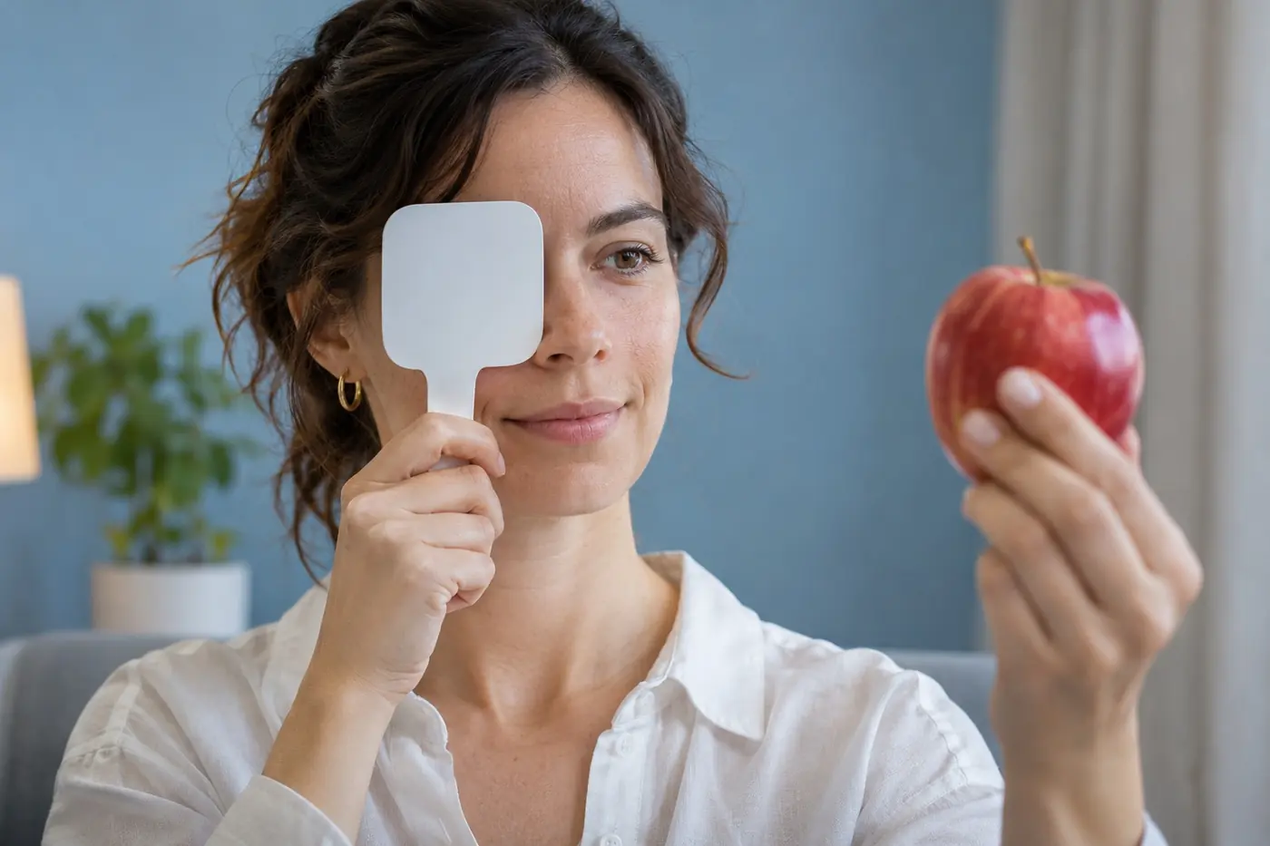
Without delaying assessment, you can look at the same uniformly coloured object under the same lighting, covering one eye then the other without pressure. Notice whether colour, brightness or clarity changes. Report the impression as it is. Screen settings, lamps, glasses and reflections can all affect perception.
A home comparison cannot reliably measure optic-nerve function. Online colour tests depend on the display and cannot exclude an acquired problem. Recognising red is not the same as seeing it with equal intensity in both eyes. Use a new difference to describe symptoms rather than to diagnose them.
A study of adults with normal eye examinations found that some reported subjective red differences between eyes. It was a limited group using a subjective test. Trying to estimate percentages or find a threshold at home is therefore unhelpful. Recent change from your own baseline, especially with other symptoms, is a stronger reason to seek assessment.
Clinic testing may include each eye’s acuity, pupil comparison, colour plates, fields, retina and optic nerve. Findings guide OCT, visual-field testing or neuro-ophthalmic investigation. One number must be interpreted alongside your experience.
Macular and optic-nerve OCT examine different structures; colour plates and fields measure different functions. An unremarkable photograph does not remove the need to investigate changed function. If MRI is required, the clinician selects the appropriate orbital or brain study. Ask which symptoms need reassessment before the next test.
Ishihara plates and online tests: what do they answer?
Familiar plates showing numbers within coloured dots mainly help identify certain red-green discrimination difficulties. They do not measure every aspect of colour perception. Reading the numbers correctly does not exclude reduced brightness, contrast or visual function in one eye.
For an acquired change, the ophthalmologist chooses tests according to symptoms and compares eyes. Colour tests may need to be combined with pupils, fields and imaging. You do not need to find a “wrong” number online to justify examination: changed perception is already clinically relevant.
Displays add variables such as brightness, night filters, colour temperature and room lighting. Different results on phone and computer may reflect those factors. Use comparisons only to describe the problem, without repeating them until reassured or delaying urgent care.
Sources · Petzold et al. – Lancet Neurology, 2022 · Hull University Teaching Hospitals NHS – Optic neuritis
All sources and further reading (9)
- National Eye Institute – Color vision deficiency
- Ministero della Salute – Ictus e TIA
- National Eye Institute – Cataracts
- National Eye Institute – Age-related macular degeneration
- American Heart Association – Retinal artery occlusion
- NHS – Temporal arteritis
- Mikolajczyk et al. – Red desaturation in healthy patients, 2022
Illustrative images from the website’s image library.
- AI-generated illustrative image
- AI-generated illustrative image